OUR EQUIPMENT
The Institute for Human Neuroscience (IHN) is a state-of-the-art cognitive neuroscience research facility that opened in 2021 and is located in Boys Town, Nebraska, a suburb of Omaha. The 25,000+ sq ft. building is quite literally the foundation of IHN. The unique layout of the building was designed by scientists with two major goals in mind: optimizing research participant experience and fostering scientific collaboration. The resulting Institute has a unique flow that unifies participant assessment with neuroimaging data collection and ultimately advanced data analytics and scientific publication, which makes IHN an ideal location for new laboratories and students seeking advanced training. The Institute for Human Neuroscience houses the latest equipment available in the field of noninvasive neuroimaging, including two Triux Neo magnetoencephalography (MEG) systems, which are the most advanced MEG units currently available, a 3T Siemens Prisma MRI system, an optically-pumped magnetometry (OPM) unit, cutting-edge brain stimulation equipment, extensive resources for high-performance computing, advanced instrumentation for movement physiology, and a new 3D printing suite. In addition, all IHN faculty, staff, and students are officed under the same roof, resulting in close collaboration and cross-fertilization of ideas.
Click on the links below to learn more about the equipment and resources available at IHN.


MEG
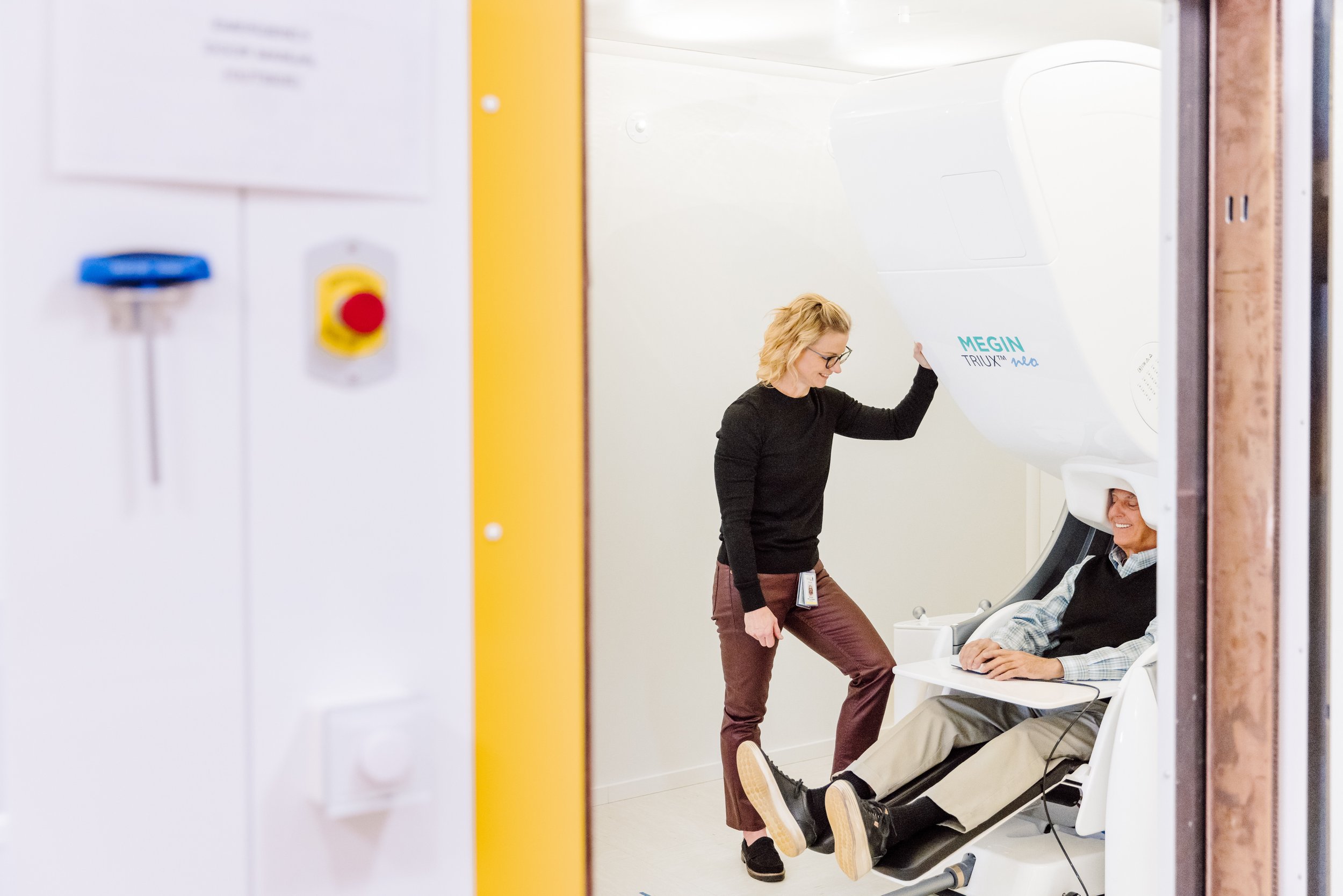
The Institute for Human Neuroscience (IHN) maintains state-of-the-art equipment for MEG imaging. In fact, IHN is the only site in the world with two MEGIN Neo MEG systems. The Neo model is the most advanced and newest MEG instrument available worldwide. It is equipped with ARMOR sensors and electronics, which provide a dynamic range that is over 300% greater than that of the Neo’s predecessor. The 306-sensor MEG array is comprised of 102 magnetometers and 204 planar-gradiometers, and each system is configured for simultaneous acquisition of 128-channel EEG. The two Neo MEG units at IHN are also equipped with an advanced fine-positioning system, which enables the nonmagnetic MEG chair to be adjusted from multiple angles to optimize the participant’s head position in the array, thereby improving signal detection and spatial precision, especially in patients. Regarding environmental noise, the instruments are housed within two-layer Vacuumschmelze magnetically shielded rooms (MSR) that are equipped with both internally- and externally driven active shielding systems (i.e., the MSR responds to environmental perturbations by generating magnetic fields to compensate). Beyond the ultra-modern “smart shields,” IHN’s two MEG facilities are equipped with the latest software for additional noise reduction. Together, these tools provide an exquisitely silent magnetic environment that is ideal for cognitive neuroscience research. The systems are also fully set up for continuous head localization and head-movement correction. The two MEG systems are configured identically, and each is equipped with a wide array of magnetically silent stimulus delivery equipment, including high-fidelity DLP video projection onto a back-projection screen using ultra-fast PROPixx presentation systems.
The Institute for Human Neuroscience at Boys Town National Research Hospital is one of only a handful of sites in the United States to have a MEG Neo system, and along with Harvard, is one of only two sites to have a Neo MEG system that was purchased using funds from the NIH S10 high-end instrumentation program. These unique multi-million-dollar systems position IHN to lead the world in research on human brain dynamics, especially regarding how development modulates neural dynamics and the impact of childhood psychiatric and neurological conditions. The combined spatial and temporal precision of MEG enables scientists to compute measures of dynamic functional connectivity, which reflect the millisecond-to-millisecond interactions among active brain regions. Such indices are among the most innovative currently available in human neuroscience.
The MEG group at IHN has been a world leader in MEG development and applications for the past decade. The MEG group is highly productive and has been the top such group since 2017, as measured by the total number of peer-reviewed publications per year. The group is also a national leader in federal funding for MEG-related human neuroscience research. Across IHN, there are more than 50 years of combined MEG experience. Many of IHN’s training and growth initiatives harness the uniqueness of the MEG program and the local expertise in neural dynamics to build on established strengths and promote innovation in the research programs of scientists across Boys Town National Research Hospital.

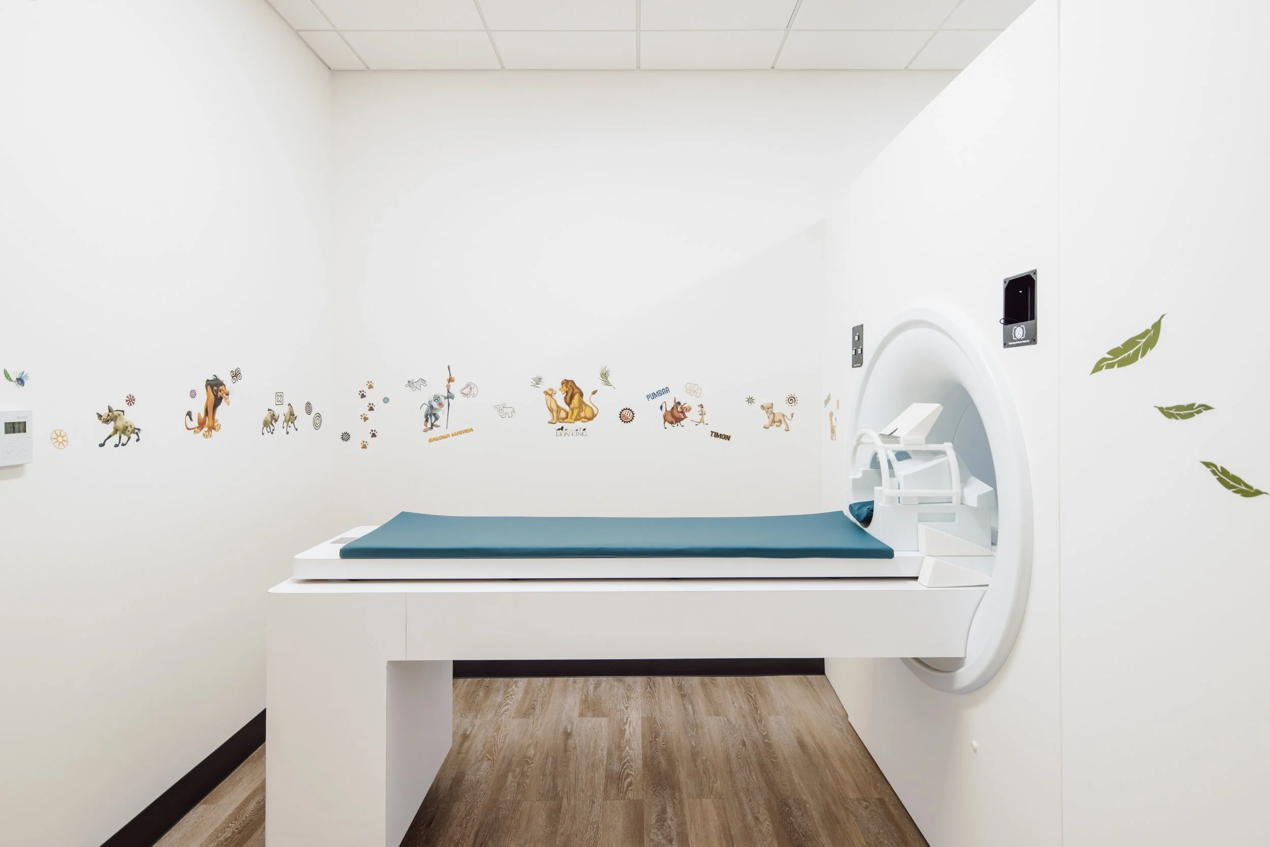
MRI
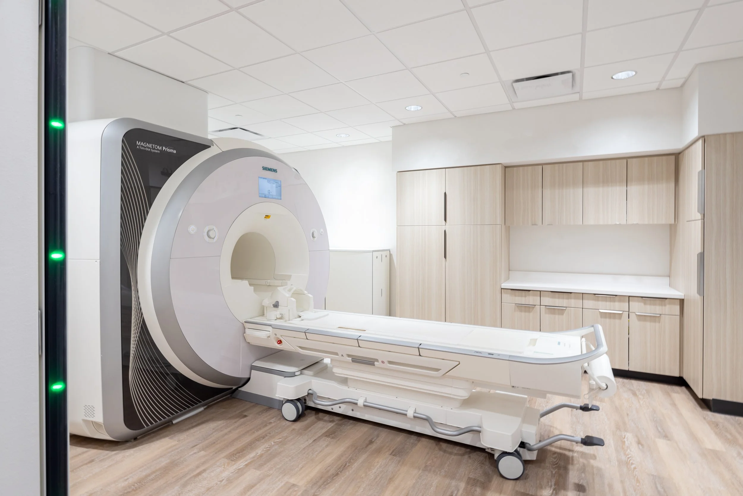
The Institute for Human Neuroscience (IHN) has a 3-Tesla Siemens Prisma MRI scanner. This research-dedicated scanner is equipped with 20-, 32-, and 64-channel head coils, as well as array of other coils for spinal cord imaging. The Prisma at IHN includes multiband imaging capabilities for advanced high-speed brain imaging, as well as all available commercial sequences offered by Siemens. These include numerous sequences for advanced diffusion, spectroscopy, structural, and functional brain imaging. Among other things, the multiband capabilities enable functional MRI volumes to be acquired in less than 300 milliseconds, whereas images of the same resolution would take about 2500 milliseconds using conventional approaches. The Prisma at IHN is also equipped with a large number of research-only, “WIP” sequences through collaborations with the CMRR at the University of Minnesota, the MRRC at the University of Pittsburgh, and the Martinos Center at MGH/Harvard. For advanced spectroscopic imaging, the scanner is equipped with the MEGA-PRESS approach for estimating local GABA concentration and the SEMI-LASER sequence for deriving estimates from hard-to-reach regions such as the amygdala. The Prisma scanner has a 60 cm bore, an ultra-fast gradient system for high-speed imaging, and 32- and 64-channel receiver coils for advanced data acquisition, permitting increased temporal resolution and reduced geometric distortions in functional MRI scans. Specifically, the system is equipped with the [204x64] XR 80/200 gradients, which are the most powerful commercially available gradients for any scanner currently on the market.
The Prisma MRI suite at IHN is also equipped with all necessary peripherals to present experimental stimuli and acquire behavioral responses. This includes a 40” in-room LCD monitor for presenting stimuli, and multiple ergonomic subject response devices, including devices that are identical to those used for the two MEG systems. The MRI suite is also equipped with the Celeritas system (Psychology Software Tools, Inc.) for syncing the scanner with stimuli and behavioral responses and a BIOPAC system for recording respiration and other vital signs for integration during fMRI data analyses.
The Institute for Human Neuroscience is additionally equipped with the in-house expertise to uniquely combine functional, structural, and diffusion-based images using the latest multimodal fusion approaches, and the data analytics wing of IHN has the necessary computing power and software for such analyses. Having both MRI and MEG technologies onsite creates unique opportunities within IHN for data fusion (i.e., fMRI+MEG) and fosters the use of current methodologies while encouraging the development of future approaches.

OPM
In early 2023, the Institute for Human Neuroscience (IHN) became the second of only two facilities in the world to install a high-density Cerca Magnetics optically-pumped magnetometer (OPM) system comprised of 208 channels. This wearable OPM system is housed in a prototype magnetically shielded room with a dense array of magnetic field-nulling coils distributed on all sides of the room. This distributed array of coils within the walls of the room enables a technique known as dynamic nulling, which effectively allows participants to ambulate during OPM recordings. Together, our OPM array and prototype room comprise one of the most advanced MEG inspired neuroimaging systems in the world.
Unlike other modern neuroimaging methods, OPM allows researchers to study the brain activity of children from birth with great spatial and temporal precision. The OPM system consists of a series of Lego-sized sensors attached to a helmet or a beanie cap in the case of infants, that can be worn by newborns, toddlers, children, and adults. Since the sensors are attached to a helmet or hat that is worn by the participant, the person can move around and interact with their environment while scientists directly record their brain activity in real-time. The latest generation of OPM systems provides full brain coverage and offers millisecond temporal resolution at a spatial precision of millimeters. Thus, the system is capable of providing the first high-resolution dynamic maps of babies and will allow scientists to understand how functions like facial recognition and language acquisition occur in the first year of life.
Scientists at IHN are also developing new helmets and other tools using the Institute’s advanced 3D Printing Core to enable the next phase of brain science discovery and the capacity to quantify nerve impulses transversing the spinal cord and other peripheral aspects of the human nervous system.

BRAIN STIMULATION
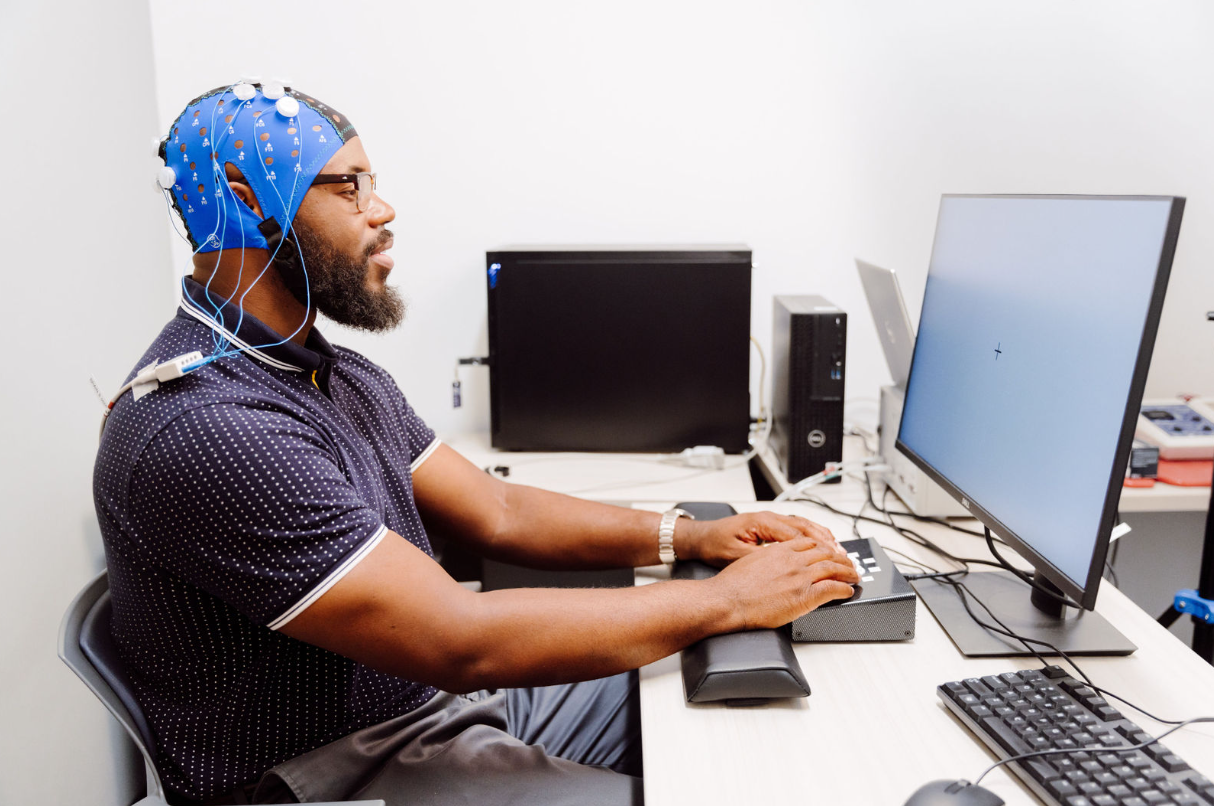
The Institute for Human Neuroscience (IHN) is equipped with state-of-the-art electrical brain stimulation equipment, including one of the few 65-lead transcranial electrical stimulation systems (tES) in existence. This Soterix 65-lead system tES system, the MxN-65 HD, went live in February 2023 and enables multi-montage approaches for precise targeting of brain regions using direct-current (tDCS), alternating-current (tACS), and other approaches. The system is also configured with the latest targeting and current density modeling software, which can be integrated with individual structural MRI data for precise targeting of specific brain regions and individualized current dosing for precision medicine applications. In addition to the 65-lead system, IHN houses a standard two-pad (tES) system, a five-lead multipolar high-definition tES system (HD-tES), and a MxN-33 lead tES system. All of these systems were developed by Soterix and can make use of the current-density and targeting software mentioned above. These systems are also equipped with settings for sham-stimulation, which allows investigators to use “placebo-controlled” experimental designs. In addition to the stimulators, there is a Polhemus digitizer for coregistering the stimulation sponges or metal electrodes to neuroanatomical images to improve spatial precision, and all users also have access to advanced software for finite-element modeling (FEM) of current flow using the participant’s individual anatomy. All of these tools are housed in a dedicated brain stimulation suite that includes a general preparation and setup area, as well as two testing/stimulation areas.

3D PRINTING SUITE
The Institute for Human Neuroscience (IHN) boasts a new, state-of-the-art 3D printing suite. This includes a Selective Laser Sintering (SLS) industrial grade 3D printer. This printer, the EOS P396, features a build volume of 340 x 340 x 600 mm and can print objects at speeds of approximately 48 mm/hour with layer thicknesses as fine as 60 microns. This SLS printer uses a powerful laser to fuse material powder (polyamide) together to create a three-dimensional object. The utilization of a laser to precisely sinter the plastic powder allows for a very high level of detail on the output printed objects. Currently, the SLS 3D Printer is primarily being used to print custom-fit OPM helmets and equipment for inside the MEG and OPM rooms.
The 1,800 square-foot 3D printing suite also includes a Raise3D Pro3 3D printer, which utilizes the Fused Deposition Modeling (FDM) method to print objects. This printer is ideal for agile and small-batch production.

HIGH PERFORMANCE COMPUTING
The Institute for Human Neuroscience (IHN) includes a dedicated computer cluster. This NIH-funded system, Orion, is comprised of four Dell PowerEdge nodes with 256 cores (Intel Xeon Turbo Platinum, 2.6 GHz, 11.2 GT/s), 2.0 TB of RAM (dual-rank, 3200 MT/s), high-speed GPUs (NVIDIA Ampere A100, 80 GB, Double Wide – Full Height), over 25 TB of high-speed data storage (Dell Enterprise, NVMe, U2, G4, P5600), and ultra-high speed networking capabilities (Broadcom 57414, dual-port, 10/25 GbE PCle). The GPUs are designed to maximize total throughput for machine learning, deep learning, and other artificial intelligence applications. The overall Orion cluster is implemented on a Rocky Linux platform with the Slurm workload manager, which helps allocate resources as jobs are setup and assigned to the cluster. Orion is physically housed in the Boys Town National Research Hospital’s Data Center, which enables tight temperature, humidity, and access control. The Data Center is linked to the Institute for Human Neuroscience through a dedicated pipe. In 2024, Orion is expected to undergo a major expansion that will add at least eight additional nodes.
The Institute for Human Neuroscience also includes a 4000+ square foot high-performance computing and data analytics space that includes seven supercomputers, each with i9 Turbo 10-core processor, 256 GB RAM, 25+ TB of high-speed disk space, and advanced graphics cards, as well as over 70 high-performance data processing workstations equipped with i9 processors, 64 GB of RAM and multiple terabytes of disk space. Each workstation is linked to a 90+ terabyte storage array for neuroimaging data and the dedicated computer cluster mentioned above. Each workstation is also configured and equipped with the latest software for advanced data analyses, including Matlab and other common software for neuroimaging and statistical analyses such as SPM, BrainStorm, FSL, AFNI, FreeSurfer, CONN, R, and other leading toolboxes. Many of the computers are also equipped with the Brain Electrical Source Analysis (BESA) software, SPSS, and current-distribution modeling software. The open-concept data analytics space encourages collaborative programming (e.g., algorithm development) and data processing efforts among students and faculty.

MOVEMENT
PHYSIOLOGY LAB
The Institute for Human Neuroscience (IHN) maintains a state-of-the-art laboratory for the biomechanical assessment of human movement. Data collected with this equipment can inform clinicians and families on what therapeutic practices could be beneficial. In turn, we hope to pinpoint personalized rehabilitative practices that will result in clinically significant improvements in the motor actions of those with developmental disabilities.
The 800-square-foot laboratory has a Vicon motion capture system and a Bertec-instrumented split-belt treadmill. The treadmill is unique in that it has force platforms under the respective belts that capture the forces applied by the legs to accelerate the center of mass during gait. The respective equipment is also used in conjunction with stimulators that can provide insights into the activity of the spinal cord, electromyographic sensors to assess changes in muscular activity, and a 64-channel electroencephalographic (EEG) to assess the role of cortical activity during gait. The lab also houses a KINARM endpoint robotic arm that is used to quantify force production, movement trajectory, sensory perceptions, and cognitive-motor decisions. The technology allows us to study how individuals adapt and learn to produce motor actions.
MOCK MRI
The Institute for Human Neuroscience (IHN) contains a full-scale Mock MRI scanner (Psychological Software Tools), which simulates the real MRI scanning environment, including the sounds and vibrations that an MRI makes during a real scan. This mock scanner is used to expose participants to the MRI environment, including the restrictive bore and loud noises, and helps reduce claustrophobia, anxiety, and body motion. The mock scanner at IHN is equipped with multiple head coils, including one that is identical to the 32-channel Siemens coil that is frequently used with the Prisma. The mock scanner is equipped with loudspeakers to play pre-recorded ambient noise (e.g., cryogenic pump) that can be mixed with real gradient noises for specific sequences (e.g., EPI for fMRI). Finally, the mock scanner is also configured with response devices (e.g., button boxes) identical to those used with the Prisma MRI system, which is interfaced with a stimulus presentation computer, head coil mirror, and a monitor located at the end of the mock bore. This allows IHN scientists to run pilot versions of their tasks prior to moving the participant into the real scanning environment, which is often helpful in pediatric and clinical populations where difficulties in understanding the task instructions are common. This powerful tool has allowed researchers at IHN to improve participant success and to collect robust MRI samples from children as young as 5 years-old.